FACILITIES
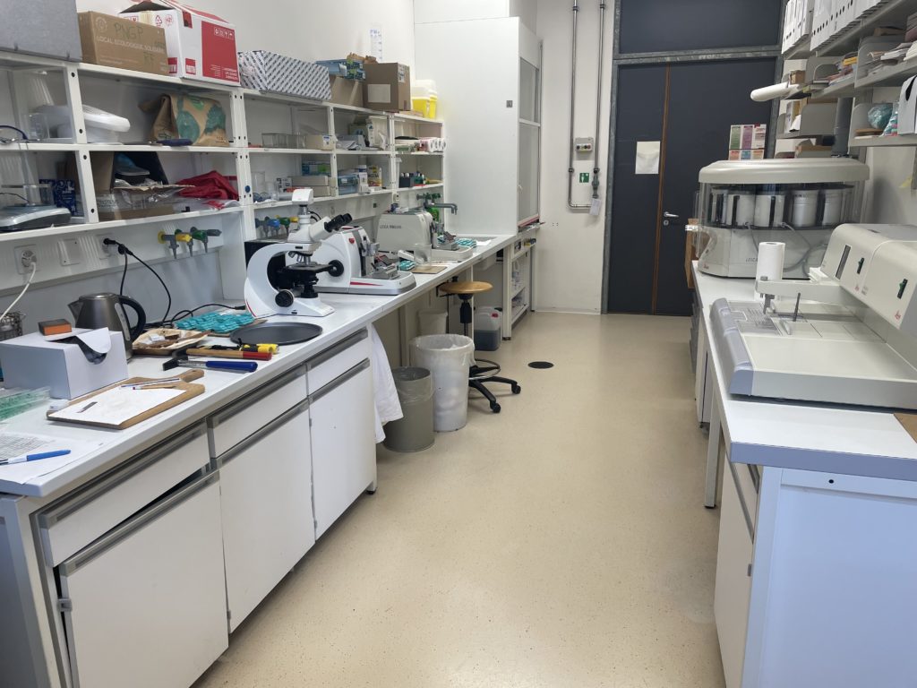
Dendrolab.ch operates fully equipped tree‑ring, wood anatomy and imaging laboratories at the University of Geneva. Our facilities cover the full workflow from field sampling to high‑resolution imaging and analysis of wood, allowing us to handle both research projects and service requests in a reliable and reproducible way.
Sample preparation
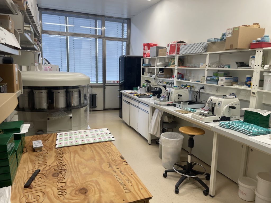
High‑quality samples are the foundation of robust tree‑ring and microstructural analyses. In our lab, we prepare samples using dedicated equipment that ensures precision and repeatability.
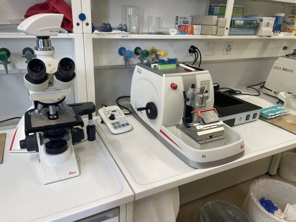
Leica rotary microtome RM 2245
Our Leica rotary microtomes RM 2245 allows the precise cutting of very thin sections from embedded wood samples. Thin sections reveal the internal structure of the material and are essential for detailed anatomical and microstructural studies. The microtome enables us to obtain uniform sections suitable for high‑resolution imaging and long‑term archiving.
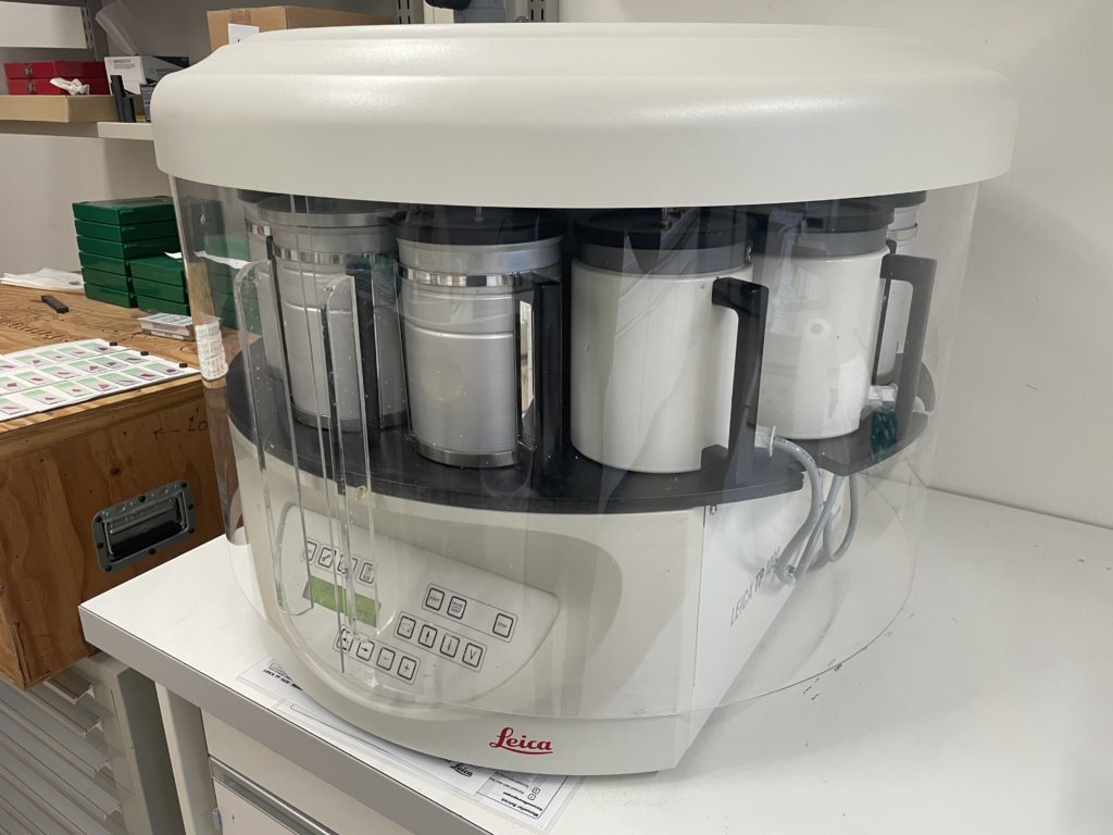
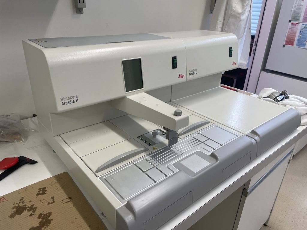
Leica sample embedding station
The Leica embedding station (LEICA TP1020 with HISTOCORE ARCADIA H-C) is used to infiltrate and embed samples in paraffin or resin before sectioning. Embedding stabilizes fragile, decayed or very small samples, including archaeological wood and delicate research material. This process improves section quality and reduces the risk of damage during cutting, which is especially important for unique or irreplaceable samples.
Together, the microtome and embedding station allow us to produce high‑quality thin sections that meet the standards required for modern tree‑ring and anatomical analyses.
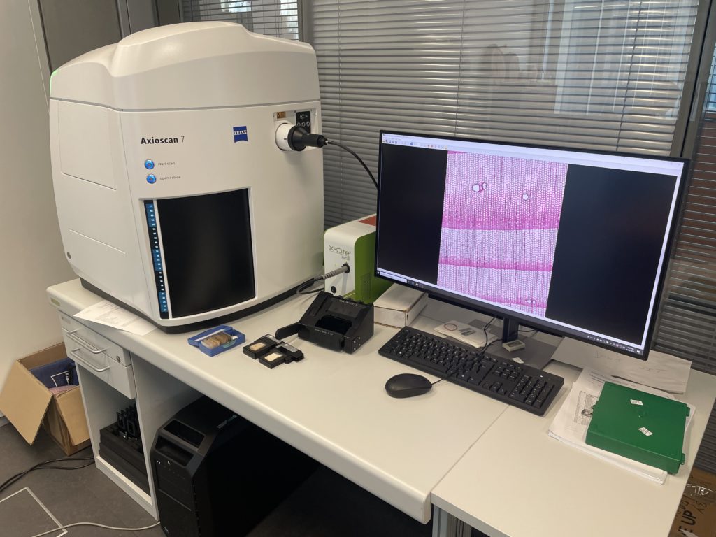
Slide scanning – Zeiss Axioscan 7
The Zeiss Axioscan 7 is an automated slide‑scanning microscope that produces high‑resolution digital “virtual slides” from brightfield or fluorescence samples. The system can handle large batches of slides and automatically focuses, tiles and stitches images to provide both an overview of the entire sample and detailed views at high magnification. These virtual slides enable quantitative measurements, remote annotation and image sharing with collaborators and clients, and they are ideal for long‑term digital archiving of valuable material.
Imaging systems Skippy and GigaMacro
Our imaging lab also includes two complementary systems that allow us to cover different spatial scales.
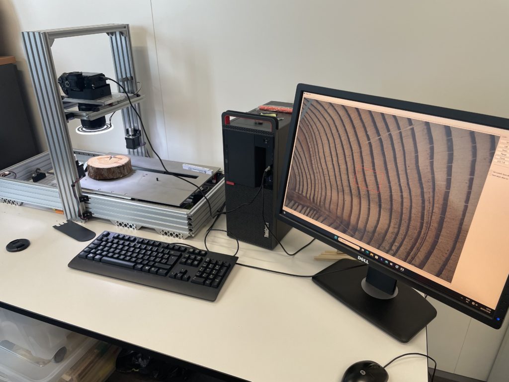
Skippy imaging system
The Skippy imaging system is used for high‑quality imaging of larger wood specimens, such as increment cores or blocks. It provides detailed visualization of tree‑ring boundaries and anatomical features and is well suited for routine dendrochronological measurements as well as more advanced anatomical investigations.
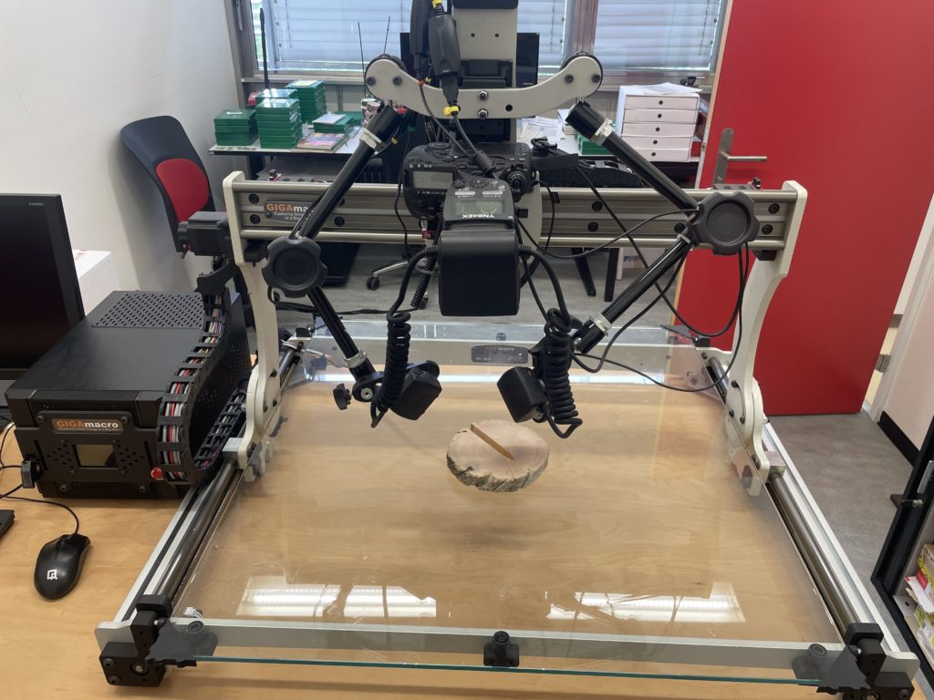
GigaMacro imaging system
The GigaMacro system is a macro‑imaging platform that produces gigapixel‑scale images of large objects or surfaces, such as polished wood discs, cross‑sections or outcrop surfaces. It allows us to capture the full extent of a sample at very high resolution, making it possible to zoom in on fine details while retaining the context of the entire specimen.
By combining the Skippy, GigaMacro and Zeiss Axioscan 7 systems, we can image samples from the scale of entire tree discs or cores down to the cellular level. This flexibility allows us to choose the most suitable approach for each project and to provide high‑quality visual material for both scientific and applied purposes.